You’ve seen it. That grainy, beige slurry in a glass jar on someone's kitchen counter. Or maybe you've looked at the back of a Fleischmann’s packet and wondered what those little dry pebbles actually look like if you zoomed in a thousand times. Looking at a picture of a yeast isn't just a biology class requirement; it’s basically looking at the engine of human civilization. We’ve been hanging out with Saccharomyces cerevisiae for thousands of years, but most people wouldn't recognize it if it hit them in the face.
It’s alive.
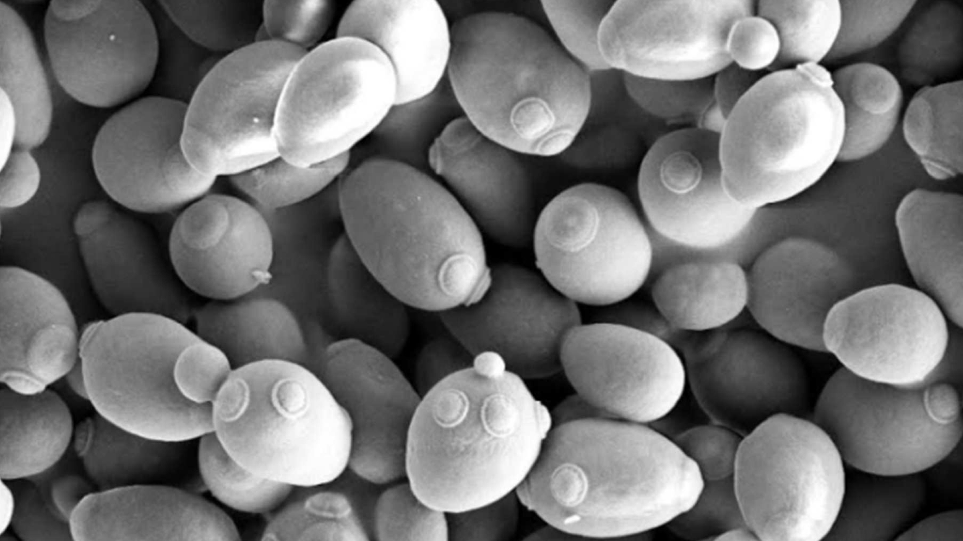
When you see a high-resolution image of yeast, you aren't looking at a plant. You aren't looking at a bacteria either. You’re looking at a single-celled fungus. It’s weirdly beautiful. Under an electron microscope, yeast cells look like smooth, egg-shaped boulders huddled together in a pile. But if you’re looking at a standard light microscope photo, they’re just tiny, translucent circles, often with little "buds" sticking off the side like a biological Mickey Mouse.
The Visual Identity of a Single Cell
If you pull up a picture of a yeast cell from a lab like the one at the University of Washington’s Department of Genome Sciences, you'll notice the "bud scars." This is the coolest part. Every time a yeast cell reproduces—which it does by quite literally pushing a new version of itself out of its own body—it leaves a permanent mark. It’s like a belly button that tells you exactly how many "kids" that cell has had.
A young cell is smooth. An old cell is covered in these circular craters.
Most people expect yeast to look green or mossy because we associate fungi with mold. Yeast is different. In its natural state, it’s creamy, off-white, or slightly tan. If you see a picture of yeast that’s bright pink or deep red, you’re likely looking at Rhodotorula, a different genus that can actually be a bit of a nuisance in your bathroom grout or on damp shower curtains.
Context matters. A photo of dry active yeast looks like a desert landscape of craggy, dehydrated spheres. Add water, and those spheres dissolve into the living, breathing cells that make your sourdough rise.
Why Microscopes Change Everything
There is a massive difference between what a baker sees and what a microbiologist sees. If you search for a picture of a yeast culture in a brewery, you’ll see "flocculation." This is just a fancy word for yeast cells clumping together and sinking to the bottom of the tank. To the naked eye, it looks like white mud.
Under a Scanning Electron Microscope (SEM), that "mud" turns into a 3D landscape. You can see the texture of the cell wall, which is made of chitin and glucans. It’s tough. It has to be. That wall is the only thing keeping the high-pressure internal environment of the cell from exploding.
Different Views for Different Needs
- Light Microscopy: This is the "classic" look. You’ll see the vacuole—a large, clear sac inside the cell that stores nutrients. It looks like a bubble inside a bubble.
- Fluorescence Imaging: Scientists use dyes to make specific parts of the yeast glow. You might see the mitochondria glowing bright green or the nucleus glowing blue. It looks like deep-space photography.
- Differential Interference Contrast (DIC): This gives the cells a 3D, shadowed look without needing a massive electron microscope. It’s how you get those "artistic" shots of yeast where they look like glass beads.
Honestly, it’s kind of wild how much we rely on these things. Every sip of beer, every slice of pizza, and even modern insulin production relies on these tiny eggs. When you see a picture of a yeast being used in a lab, it’s often because they are using it as a "model organism." Since yeast cells have a nucleus just like human cells do, scientists like those at the Nobel-prize-winning Nurse lab have used them to figure out how cancer works.
The Sourdough Obsession
Lately, the most common way people interact with yeast imagery is through the lens of fermentation. A picture of a yeast starter (levain) is a mess of bubbles. Those bubbles aren't the yeast itself. They are the carbon dioxide gas trapped in a gluten network—the "exhale" of the yeast eating flour sugars.
If you’re trying to diagnose your starter at home, the visual cues are everything.
A healthy starter has a frothy, mousse-like texture. If the "picture" you’re looking at in your kitchen involves a layer of dark liquid on top, don't panic. That’s "hooch," a byproduct of fermentation that tells you your yeast is hungry. If the yeast has turned fuzzy or orange, that's when you toss it. True Saccharomyces doesn't do fuzz.
Visualizing the invisible
We often forget that yeast is everywhere. It’s on the skin of grapes. It’s in the air. It’s even on you. When you look at a picture of a yeast found on a wine grape, it looks like a dusting of fine white powder. This is the "bloom." Winemakers in regions like Burgundy often rely on this "wild" yeast rather than adding store-bought packets. They trust the visual presence of that white dust to ferment an entire vintage.
It’s a gamble, but it’s one humans have been taking since the Egyptians.
Taking Action: How to Observe Yeast Yourself
You don't need a $10,000 Leica microscope to see this stuff. If you have a decent hobbyist microscope (even a $100 one), you can create your own picture of a yeast colony at home.
- The Sugar Hack: Mix a pinch of dry yeast with warm water and a teaspoon of sugar. Let it sit for ten minutes until it gets foamy.
- The Slide Prep: Take a tiny drop of that foam—just a tiny bit—and put it on a glass slide. If the liquid is too thick, you won't see individual cells; you'll just see a blur.
- Staining: Use a drop of Methylene Blue if you can get it. This is a "vital stain." Dead yeast cells will turn dark blue because their damaged membranes can't pump the dye out. Living cells will stay clear or very pale. This is literally how commercial brewers check the health of their "pitch."
- Magnification: Start at 400x. At this level, you’ll see the "budding" clearly. You’ll see the tiny daughter cells clinging to the mother cells.
By understanding the visual cues of yeast—from the microscopic bud scars to the macroscopic bubbles in a bowl of dough—you get a better grip on the fermentation process. Stop thinking of it as a powder. Start thinking of it as a massive population of tiny, egg-shaped organisms that are constantly eating, breathing, and reproducing. When you look at a picture of a yeast next time, look for the scars. They tell the story of a cell that has already worked hard to make something delicious.